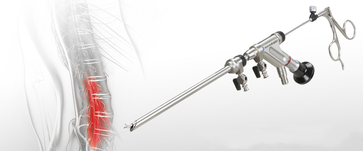
Endoscopic discectomy
is a minimally invasive surgical procedure used to remove herniated disc material that is causing pain in the lower back and legs (lumbar), mid back (thoracic), or neck and arms (cervical).
Endoscopic discectomy is the least invasive and most effective surgical technique for treating spinal disc herniation patients. With endoscopic spine surgery, surgeons do not need to remove bones and muscles in order to remove herniated discs. Surgeons can see the spine with a camera, smaller than a smart phone camera, through a small surgical port (tube). Large incisions are avoided. The procedure does not traumatize your spine like traditional spine surgeries do. The whole procedure for a disc herniation takes about 30 minutes. The patient goes home in 2-3 hours when the surgery is done.
Endoscopic Spine Decompression
An endoscopic micro-invasive decompression is the least invasive technique that allows direct visualization of the disc and the nerves. This procedure is used for decompressing nerve roots damaged by narrowing of the spinal canal and foramens caused by bulging disc, herniated discs, facet arthritis, bone spur, scoliosis, and spondylolisthesis
It is usually indicated in patients with neurological symptoms such as radiating pain down the legs, tingling, numbness, weakness, difficulty breathing, and in some cases bowel/bladder problems. Most of our patients have not found adequate relief with non-surgical modalities including but not limited to pain management injections or conservative treatments. This procedure can also help in relieving pain associated with spinal stenosis and low back arthritis.
What is an endoscopic decompression?
An endoscopic decompression is the least invasive surgery that allows the surgeon to decompress the spinal nerves in the spinal canal with less than 1 cam incision. The surgery is utilizing endoscope, real time EMG, real time radiographic images so the surgery is very safe and effective
Many patients who suffer from sciatica, referred pain down either legs, and/or low back pain may be a candidate for this procedure. This procedure can also help in relieving pain associated with spinal stenosis and low back arthritis.
Benefits associated with a Micro-invasive endoscopic decompression
- Least invasive technique – less trauma to muscles and soft tissue than with traditional open surgery
- Fast recovery time
- Minimal pain or discomfort following the surgery
- Immediate leg pain relief in most cases
- Fewer complications and risks than open spine surgery
- Small incision and minimal scar tissue
- High success rate and sustained success of the therapy
- No or minimal blood loss
- No hardware placement or loss of mobility
Endoscopic Facet joint rhizotomy
Endoscopic rhizotomy allows the spine surgeon to place a small cannula and an endoscope attached to an HD camera, inside the patient’s back and target visually the medial branch nerve. A radiofrequency probe is used through the endoscope to ablate the medial branch nerve. Because the endoscope allows us to see the medial branch nerves that cause low back pain, we can ablate and sever these nerves with certainty. Also, the endoscopic rhizotomy surgery provides much longer pain relief (up to 5 years) to patients than the traditional pain management RFA. This procedure has spared many patients of more invasive spine surgery such as a spinal fusion. patients have been very satisfied with the results and typically go back to work or an active lifestyle in a few weeks.
The advantage of the endoscopic rhizotomy is that it is often used when other surgeons have recommended no treatment at all or spinal fusion for the back. The procedure is less invasive and a great alternative for patients suffering from debilitating back pain and spasms.
- High success rates, 90% or above
- Utilizes an HD camera coupled to an endoscope which provides the physician a superior view to that of traditional surgical techniques
- No spinal fusion is necessary thus preserving the spinal column and the disc
- Less than i cm incision minimizes potential skin scarring
- No muscle or tissue tearing thus less scar tissue and preserve spinal mobility
- No significant blood loss
- Conscious sedation reduces the risk associated with general anesthesia
- Less post-operative pain and need for narcotic medicines
- Less recovery time needed
- Return to work sooner
What Conditions Does Endoscopic Facet Rhizotomy Treat?
- Facet joint syndrome
- Facet related arthritis
- Chronic low back pain
- Back spasms related to the facet joint
- Failed back surgery
Endoscopic sacroiliac joint rhizotomy
The sacroiliac joint can become inflamed and painful or injured for a number of different reasons.it is one of the most common causes of lower back pain, as in facet joint rhizotomy we utilize a small endoscope through a 5 mm incision and insert a radiofrequency probe through the scope in order to burn the sensory nerves supplying sensation to the Sacroiliac joints. The endoscope allows us to see the lateral branches (sensory nerves )and ablate them. patients have been very satisfied with the results and typically go back to work or an active lifestyle in a few weeks, this allows up to a five year pain free period. It also has superior results over thermal ablation by needles and SIJ steroid injections.
Endoscopic Coccygeal nerves rhizotomy
Treatment of chronic coccyx related pain can often be difficult, especially when it comes to long term improvement. As with other pain management options, coccyx treatments can be temporary in that the goal is to alleviate the pain while the body attempts to heal itself. If the coccyx pain has become chronic, meaning that it has been persisting for over 6 months, it is less likely to get better and longer term treatment options have been less likely to improve the condition. Coccygectomy is no longer a good solution for coccygeal fractures, it carries major risks and complications as well.
The endoscopic radiofrequency ablation of the coccygeal nerves involves using an endoscope (small tube) through which the coccyx and its nerve supply can be directly visualized. Through the endoscope one can then insert small tools and then ablate/burn the nerves under direct visualization. The endoscope itself is similar to the small scope commonly used for arthroscopic surgery. Skin incision size is typically 0.5 cm long (less than ¼ inch) and requires only a small stitch under the skin to close.


